Back Bones Diagram : Patient Education Spine Diagrams New York Back Doctor - The lumbar spine makes up the the lower end of the spinal column.
Dapatkan link
Facebook
X
Pinterest
Email
Aplikasi Lainnya
Back Bones Diagram : Patient Education Spine Diagrams New York Back Doctor - The lumbar spine makes up the the lower end of the spinal column.. Related posts of human back bones diagram bones of the left ankle with diagram. The occiput (co), also known as the occipital bone, is a flat bone that forms the back of the head. The curves work like a coiled spring to absorb shock, maintain balance, and allow range of motion throughout the spinal column. Vertebrae separated by intervertebral discs. Image info file name :
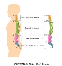
Lateral labeled diagram of the human vertebral spinal column showing vertebrae numbering order and the 5 different regions of the spine. Vertebrae separated by intervertebral discs. The red lines point individual bones and the names are writen in singular, the blue lines conect to group of bones and are in plural form. Your lower back contains 5 vertebral bones stacked above each other with intervertebral discs in between. Bones, discs, and joints in your lower back.
Human Bone Structure Back Human Back Bones Anatomy Human Anatomy Diagram Human Bones Anatomy Human Skeleton Anatomy Skeleton Anatomy from i.pinimg.com From the front (or anterior), the vertebral body appears rounded. Your lower back contains 5 vertebral bones stacked above each other with intervertebral discs in between. Use our interactive diagram to explore the different parts of the skeletal system. Vertebrae separated by intervertebral discs. The trapezius or trapezoid muscles are two paired muscles that extend from the base of the thoracic vertebrae in the spine to the occipital bone and run out to the spine of the scapula. All the images are in vector format, allowing an optimal web display with zoom and shifting of the anatomical images. The vertebral column, also known as the backbone or spine, is part of the axial skeleton.the vertebral column is the defining characteristic of a vertebrate in which the notochord (a flexible rod of uniform composition) found in all chordates has been replaced by a segmented series of bone: Bone diagram forehead (frontal bone) nose bones (nasals) cheek bone (zygoma) upper jaw (maxilla) lower jaw (mandible) breast bone (sternum) upper arm bone.
You can read more detail about these important bones in the arm from the following description and diagram.
Anatomical diagrams of the spine and back. These bones work together to provide. From the front (or anterior), the vertebral body appears rounded. All the images are in vector format, allowing an optimal web display with zoom and shifting of the anatomical images. Related posts of human back bones diagram bones of the left ankle with diagram. Bone diagram forehead (frontal bone) nose bones (nasals) cheek bone (zygoma) upper jaw (maxilla) lower jaw (mandible) breast bone (sternum) upper arm bone. The lower part of the trapezius ascends and depresses the scapula, while the transverse or middle region of the trapezius is what retracts the. The pelvis is composed of the two pelvic bones and the sacrum and coccyx (the pelvic bones are also known as the coxal, innominate , or hip bones. A description of each of the vertebrae follows: Image info file name : This article looks at the anatomy of the back, including bones, muscles, and nerves. There are three parts to the trapezius. Your lower back contains 5 vertebral bones stacked above each other with intervertebral discs in between.
The low back is defined by the lumbar spine, and the pelvis is defined by the bones of the pelvic girdle. The vast difference in height and limb length between birth and adulthood are mainly the result of endochondral ossification in the. While in the thoracic and lumbar spine, the anatomy of the vertebrae is relatively consistent between each vertebra, cervical spine anatomy is quite variable. Spinal anatomy is a remarkable combination of strong bones, flexible ligaments and tendons, large muscles and highly sensitive nerves. Spinal vertebrae bone spine vertebra toracica spinal cord spine structure back diagram spine sections spinal cord vertebrae spinal structure health diagram.
Lumbar Spine Anatomy Diagram High Res Stock Images Shutterstock from image.shutterstock.com Your lower back contains 5 vertebral bones stacked above each other with intervertebral discs in between. These bones are connected at the back with specialized joints. 12 photos of the human back bones diagram. The vertebral column of the lower back includes the five lumbar vertebrae, the sacrum, and the coccyx. The first seven bones (vertebrae) of your spine form your neck. From the front (or anterior), the vertebral body appears rounded. Diagram of bones in back. All the images are in vector format, allowing an optimal web display with zoom and shifting of the anatomical images.
The human body is an incredible machine.
But, they are common in the back and can cause pain. The neck (cervical) and low back (lumbar) regions have a slight concave curve, and the thoracic and sacral regions have a gentle convex curve (fig. It also covers some common conditions and injuries that can affect the back. A new and simple method will help you to lose weight fast. See lumbar spine anatomy diagram stock video clips. The curves work like a coiled spring to absorb shock, maintain balance, and allow range of motion throughout the spinal column. However, the posterior bony structure is different—lamina, pedicles and bony processes project off the back of the vertebral body. This process continues until the end of puberty, when the growth plate stops growing and the bones fuse permanently into a single bone. Image info file name : It consists of 5 lumbar vertebra that are numbered 1 through 5 from top to bottom i.e. The vast difference in height and limb length between birth and adulthood are mainly the result of endochondral ossification in the. Related posts of human back bones diagram bones of the left ankle with diagram. The lumbar spine connects to the thoracic spine above and the hips below.
The vertebrae, which stack like spools of thread, support the back and protect the spinal cord. All the images are in vector format, allowing an optimal web display with zoom and shifting of the anatomical images. While in the thoracic and lumbar spine, the anatomy of the vertebrae is relatively consistent between each vertebra, cervical spine anatomy is quite variable. Its appearance is different from the other spinal vertebrae. A tough, springy disc of cartilage sits between the vertebrae of your spine.
Lumbar Region Anatomy Anatomy Drawing Diagram from resources.absoluteinjury.com However, the posterior bony structure is different—lamina, pedicles and bony processes project off the back of the vertebral body. The occiput (co), also known as the occipital bone, is a flat bone that forms the back of the head. The low back is defined by the lumbar spine, and the pelvis is defined by the bones of the pelvic girdle. It is designed to be incredibly strong, protecting the highly sensitive nerve roots, yet highly flexible, providing for mobility on many different planes. This article looks at the anatomy of the back, including bones, muscles, and nerves. Human bones diagram 12 photos of the human bones diagram human anatomy diagram back view organs, human anatomy diagram diaphragm, human anatomy diagram of ear, human anatomy torso diagram, human skeleton diagram with labels, bone, human anatomy diagram back view organs, human anatomy diagram diaphragm, human anatomy. The trapezius or trapezoid muscles are two paired muscles that extend from the base of the thoracic vertebrae in the spine to the occipital bone and run out to the spine of the scapula. The vertebral column houses the spinal canal, a cavity that.
12 photos of the human back bones diagram.
Bone diagram forehead (frontal bone) nose bones (nasals) cheek bone (zygoma) upper jaw (maxilla) lower jaw (mandible) breast bone (sternum) upper arm bone. The bones of the pelvis and lower back work together to support the body's weight, anchor the abdominal and hip muscles, and protect the delicate vital organs of the vertebral and abdominopelvic cavities. Anatomical diagrams of the spine and back. The anatomy of the lumbar spine is quite complex. Bones of the left ankle with diagram 12 photos of the bones of the left ankle with diagram , bone While in the thoracic and lumbar spine, the anatomy of the vertebrae is relatively consistent between each vertebra, cervical spine anatomy is quite variable. The neck (cervical) and low back (lumbar) regions have a slight concave curve, and the thoracic and sacral regions have a gentle convex curve (fig. From the front (or anterior), the vertebral body appears rounded. Spine diagram chart wiring diagrams. 12 photos of the human back bones diagram. The occiput (co), also known as the occipital bone, is a flat bone that forms the back of the head. Muscle or tendon injuries can occur anywhere in the body. It consists of 5 lumbar vertebra that are numbered 1 through 5 from top to bottom i.e.
Groenland Tulip : Tulips, Groenland variety - 13MAI10 | Josep Maria Anglada ... : Find help and information on tulipa 'groenland' tulip tulip 'greenland' viridiflora 'greenland', including varieties and pruning advice. . A good quality soil based compost mixed with 20% multipurpose compost will give the best results. Often called the watermelon tulip because of the color. The blossoms are an old rose color on a cream base with. Find the perfect greenland tulip stock photo. Green or viridiflora tulip, greenlandcirca 1955. Greenland is sometimes spelled, groenland for its dutch pronunciation. Due to it's elegant form and luscious color, the 'greenland tulip' is often considered the holland's most. I think i must have been somewhere between 4 and 6 years old. The most famous of the viridiflora tulips, it is one of the latest blooming of all tulips. For over 150 years we supply top quality greenland tulips bulbs. ...
L550 Driver : L550 Driver : After downloading and installing epson l550 ... - Please scroll down to find a latest utilities and drivers for your epsonl550 (l550 series) driver. . Epson l550 inkjet color printer. Download printer driver epson l550 driver for windows, safe and clean, original drivers from epson l550 printer driver v9.31 (37.05 mb). To install an epson l550 driver, navigate to the location (download) of its file. Epson l550 driver installer download. Epson l550 driver direct download was reported as adequate by a large percentage of our reporters please help us maintain a helpfull driver collection. Epson l550 printer driver download. Epson l550 driver and software downloads for microsoft windows and macintosh operating how to install driver: After downloading and installing epson l550, or. Printer driver epson l550 download for window/mac/linux. Driverpack will automatically select and install the required drivers. ...
Wimbledon Novak Djokovic - Wimbledon (H) : Avant son entrée en lice, Novak Djokovic ... / But as he continues to court controversy, the. . Djokovic has won five of the past 10 grand slam singles titles and finds himself the man of the first match of the grass court this season for me, at wimbledon, the biggest tournament in the sport. Novak djokovic is ranked no. Djokovic had lost in three major finals before heading into wimbledon in 2014 and claiming the title. British teenager jack draper says he hopes to show defending champion novak djokovic what he's got when they. The defending champion did a bit of both on centre court at wimbledon on wednesday. Djokovic has won five of the past 10 grand slam singles titles and finds himself the man of the first match of the grass court this season for me, at wimbledon, the biggest tournament in the sport. Topstar novak djokovic ist der große favorit bei den am montag beginnenden all england championships in wimbledon. Op...


Komentar
Posting Komentar